
MS Lesion Segmentation Research
Overview
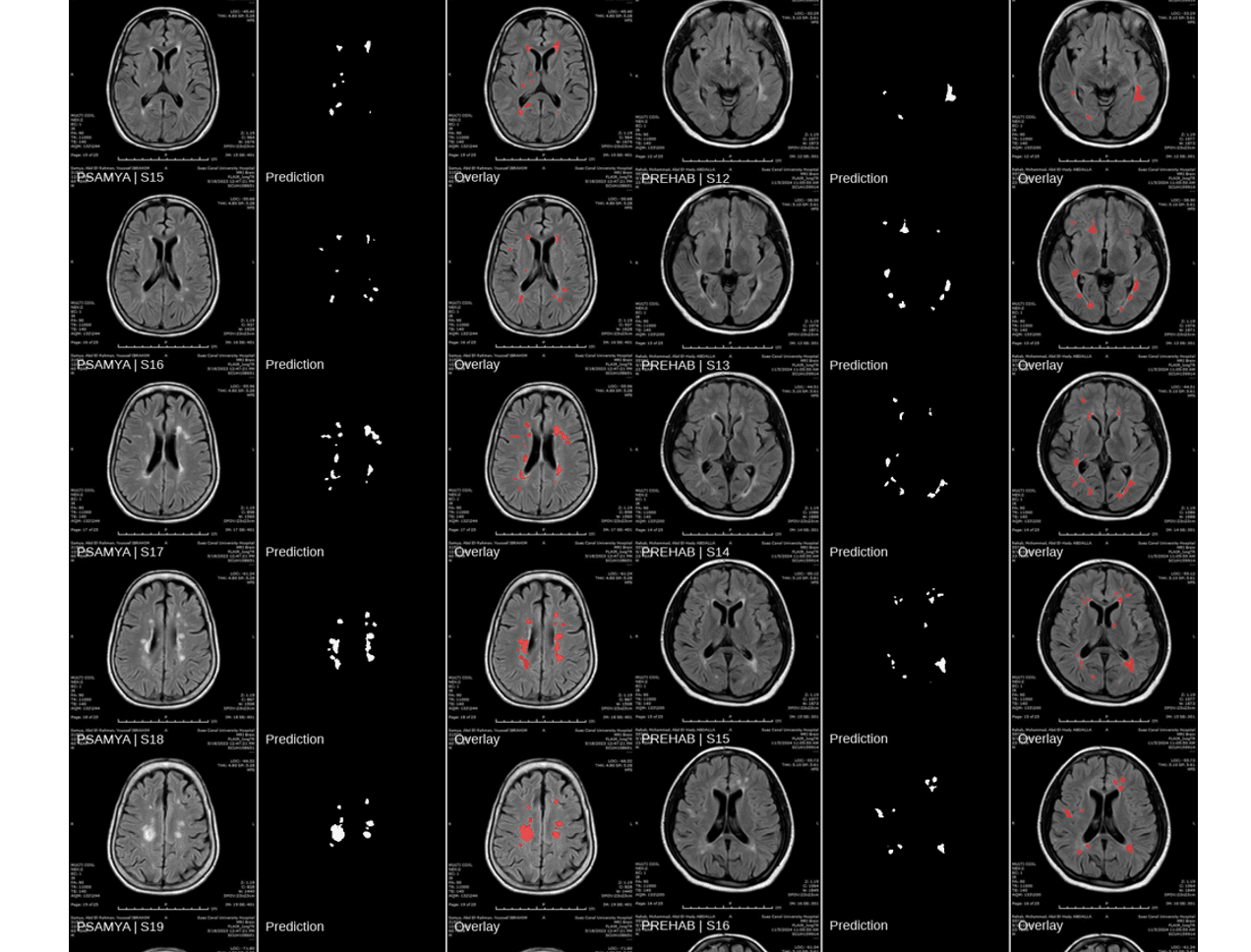
This research project is a collaboration between the Faculty of Medicine and Faculty of Engineering at Suez Canal University, focused on automated multiple sclerosis lesion segmentation in brain MRI. I contributed to the development and evaluation of U-Net-based deep learning models using 2D MRI slices derived from 3D volumes, combining public datasets with a clinically labeled dataset.
The project progressed through more than 40 controlled experimental phases, systematically evaluating changes to model architecture, loss functions, and data configuration. Each phase was isolated and logged to determine which changes produced measurable improvements rather than attributing performance gains to multiple simultaneous modifications.
Challenges & Solutions
Converting 3D MRI volumes into 2D slices introduced a risk of patient-level data leakage if slices from the same patient appeared in both training and evaluation sets.
Split the dataset by patient rather than by individual slice, ensuring no single patient's data appeared in both sets.
With many candidate changes to architecture, loss functions, and data configuration, it was difficult to know which changes were actually responsible for performance gains.
Conducted controlled ablation experiments across 40+ phases, changing one variable at a time and maintaining structured records of each configuration, result, and experimental rationale.
Clinical background as a physician was applied directly to the dataset preparation and annotation process, working alongside radiologists to review and label MRI lesion data and ensure that ground-truth annotations met both medical and engineering accuracy standards.
AI Research Collaborator